

 Eye disorders in dogs
Eye problems in dogs can stem from various causes such as hereditary factors, aging, immune responses, metabolic imbalances, infections, or injuries following accidents.
These issues may affect any part of the eye, its surrounding tissue, or the eyelids, often leading to discomfort, vision loss, and disruption of normal activities for your dog, impacting their overall quality of life.
Eye problems can arise at any phase of a dog’s life. While specific breeds might be more prone to certain conditions, no breed is entirely free from the risk of eye issues.
Early identification and prompt treatment play a crucial role in preserving your dog’s comfort, appearance, and eyesight in many conditions.
1. Cataracts in dogs
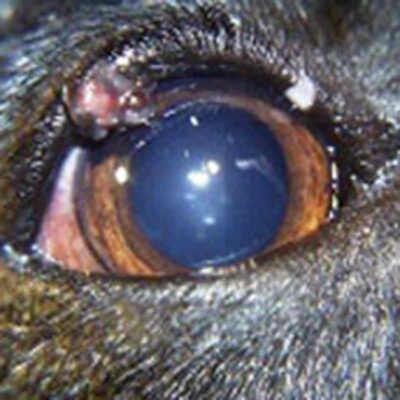
A cataract is a condition where the lens of the eye becomes cloudy, resulting in blurred vision in humans, dogs, and other animals. Since cataracts can progress to glaucoma and eventual blindness, it’s crucial to diagnose and track their development.
Causes of cataracts in dogs
While in humans cataracts often indicate aging, in dogs, this isn’t always the case. Cataracts may emerge due to various factors such as diseases like diabetes, inflammation in the eye (uveitis), aging, eye injuries, or they can be inherited.
Understanding your dog’s complete family medical history can be beneficial, especially if there’s a higher risk of cataract development.
Common triggers for cataracts in dogs:
- Congenital (present from birth)
- Diabetes mellitus
- Electric shock
- Exposure to radiation or harmful substances
- Eye injuries
- Glaucoma
- Inherited traits in specific breeds
- Lens dislocation
- Nutritional imbalances, including low calcium
- Persistent uveitis (eye inflammation)
Some dog breeds have a higher likelihood of developing cataracts, such as:
- American cocker spaniels
- Bichon Frises
- Boston, silky, and smooth fox terriers
- Golden Retrievers
- Miniature and standard Poodles
- Miniature Schnauzers
Spotting the signs of cataracts in your dog
As your dog ages, you may notice a cloudy or gray-blue appearance in their eyes. This often common condition is called nuclear sclerosis, a result of the natural aging process of the lens. It doesn’t affect vision or cause discomfort. However, nuclear sclerosis can resemble cataracts. The best way to differentiate between the two is to schedule a vet check-up for your dog.
It’s important not to delay this vet visit because cataracts are progressive, and an early diagnosis might help halt the advancement of the condition. If your dog has diabetes, additional signs of potential cataract development may include increased thirst, more frequent urination, and weight loss
Your veterinarian will conduct a comprehensive yet straightforward examination of your dog’s eyes. If they suspect cataract development, they will direct you to a veterinary ophthalmologist. The specialist can assess the extent of the condition and suggest strategies for monitoring and addressing the issue.
Treatment of cataracts in dogs
Advancements in veterinary medicine offer promising outcomes for dogs affected by cataracts. Your veterinarian might opt for specialized eye drops to address eye inflammation. However, if your dog has already experienced vision loss due to cataracts, surgery could be considered.
Veterinary ophthalmologists possess the ability to potentially restore lost vision through surgery. This surgical procedure involves replacing the affected lens with an acrylic or plastic alternative.
Cataract surgery demands meticulous post-operative care, but it boasts a notably high success rate. Your veterinarian may suggest this surgery if they believe it can aid in restoring your dog’s eyesight.
2. Collie Eye Anomaly (CEA)
Collie eye anomaly (CEA) is a condition characterized by the abnormal development of the eye in dogs, primarily affecting breeds like the Collie, Shetland Sheepdog, Australian Shepherd, and Border Collie. In certain regions, it is estimated that up to 75 percent of Collies may be affected by this disorder.
In its mildest form, CEA induces minor alterations in the choroid, the vascular layer at the back of the eye, with minimal impact on vision. CEA can impact various parts of the eye, including the retina, choroid, and, in severe cases, the sclera and optic nerve.
Being an inherited disease, most eye lesions associated with CEA are present at birth. Detecting even minor lesions can be challenging after three months of age, necessitating early examination at six to seven weeks for collies intended for breeding programs. It’s crucial to avoid breeding dogs with even minor lesions, as their offspring may inherit more severe forms of the disorder.
CEA follows a simple recessive pattern, where affected animals possess two CEA genes, implying that both parents are either affected dogs or carriers.
Diagnosing CEA is most effective between six and 12 weeks of age. The condition is identified by observing specific eye lesions, and the primary diagnostic tool is indirect funduscopy. This involves examining the retina through the pupil using a lens and ophthalmoscope, providing valuable insights into the presence and extent of CEA in affected dogs.